education | vision   
about | home
 

Transplant Nephrectomy Case Opinion

Submitted by Dr. Ian Gibson


We have had a case of sudden graft loss after over 2 years post-transplant, with an unusual histology.  I would be interested in your thoughts.

The clinical history is a 44 year old male, ESRD due to reflux nephropathy, first cadaveric renal transplant 1999 with good function.  He recently had moderately severe hypertension, and ACE inhibitor therapy was started.  On 3rd January 2002, he was seen at a clinic obviously unwell, with sudden loss of function, anuric, sCr had increased from 170 to 440umol/l and febrile.  Perfusion scan showed renal artery occlusion.  No biopsy was performed; he had a transplant nephrectomy the next day.

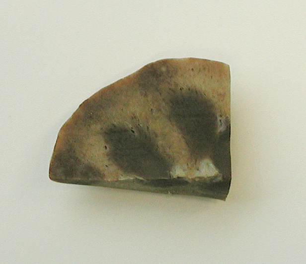
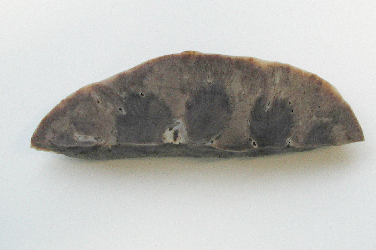
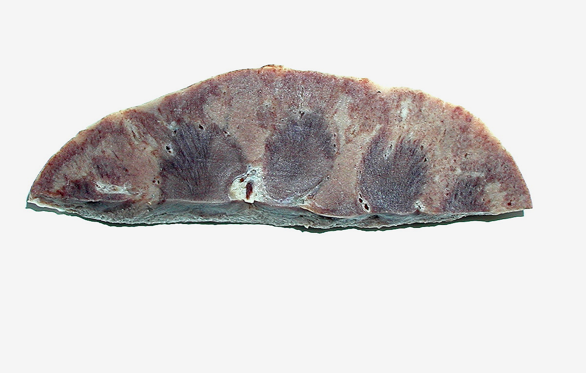
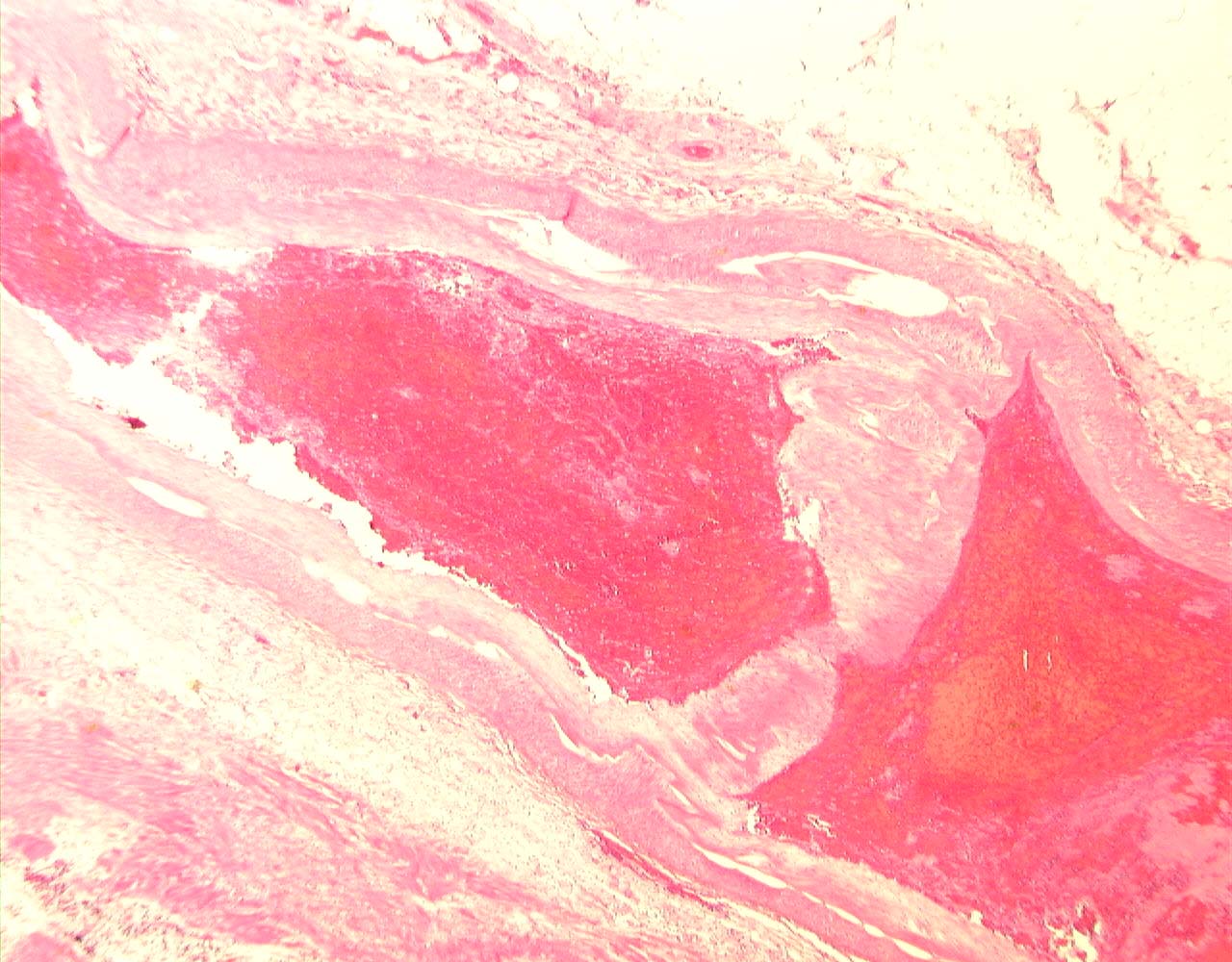
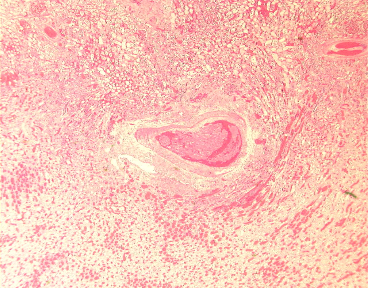
The kidney, on slicing, showed diffuse cortical pallor and multifocal paler cortical areas.  The main renal artery appeared thrombosed.


(To View Original Images Click on Thumbnail)


Gross_kidney_Gibson_1.jpg (280588 bytes) Gross_kidney_Gibson_5.jpg (969069 bytes) Gross_kidney_Gibson_7.jpg (443608 bytes)

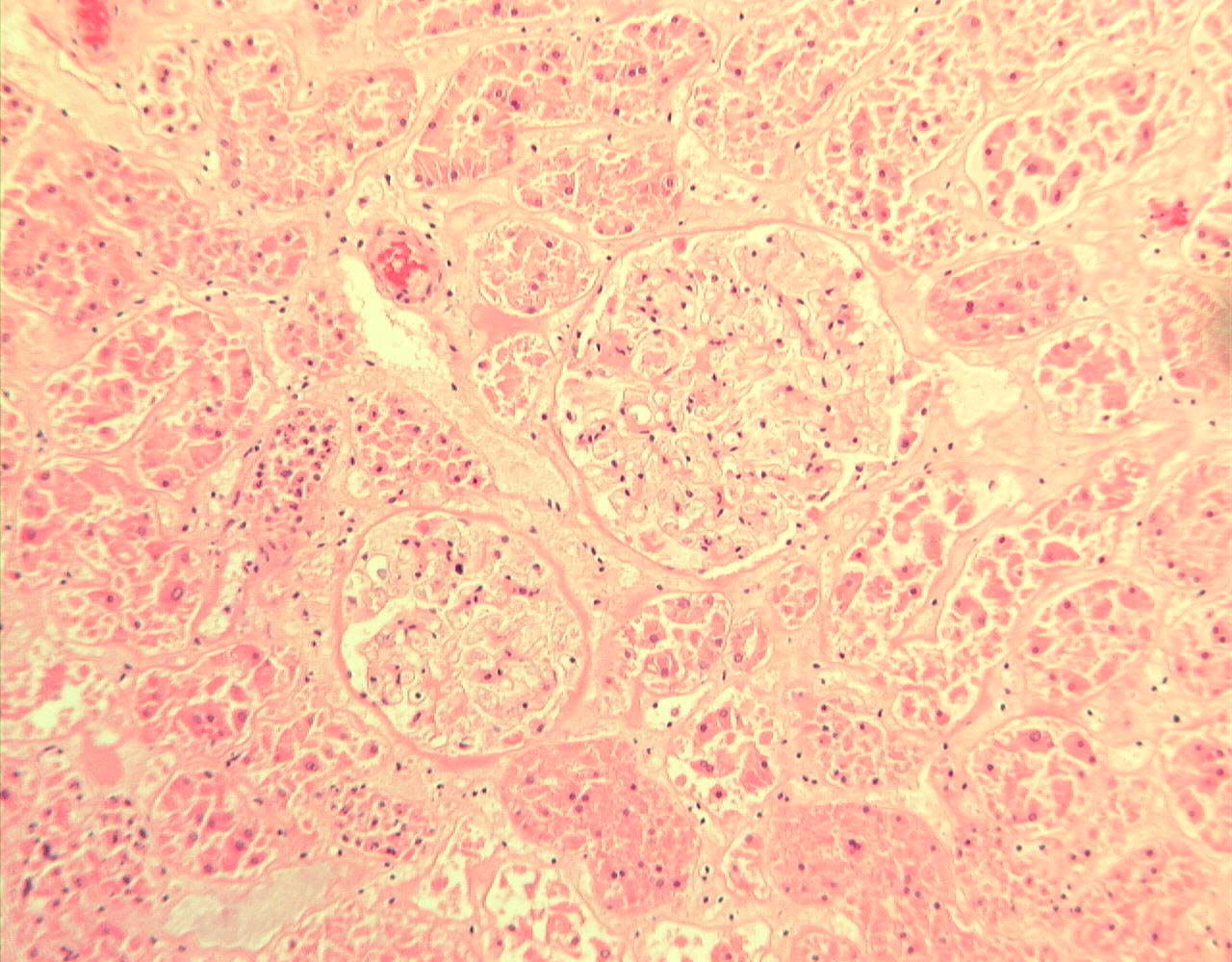
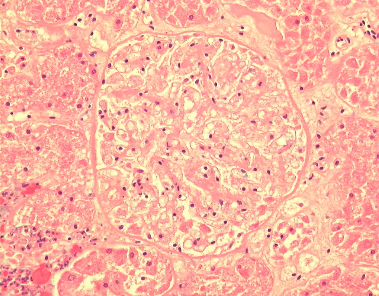
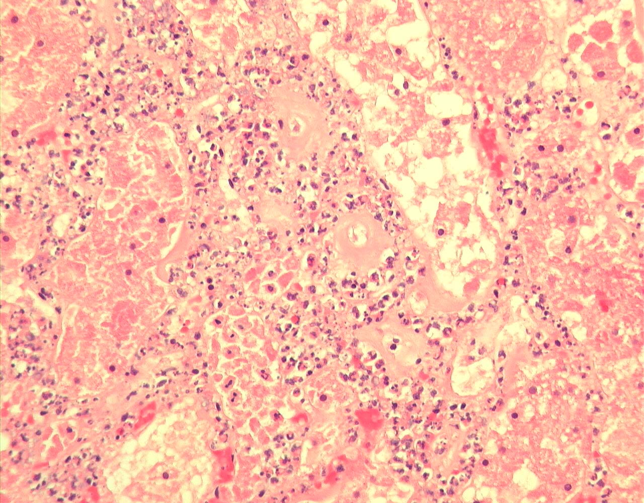
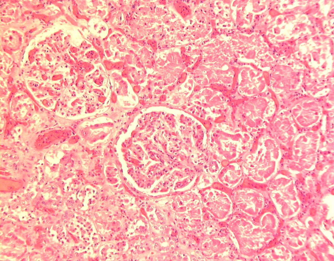
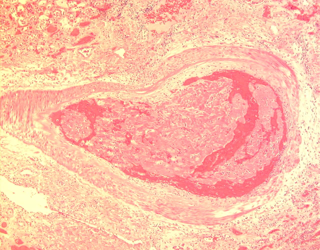
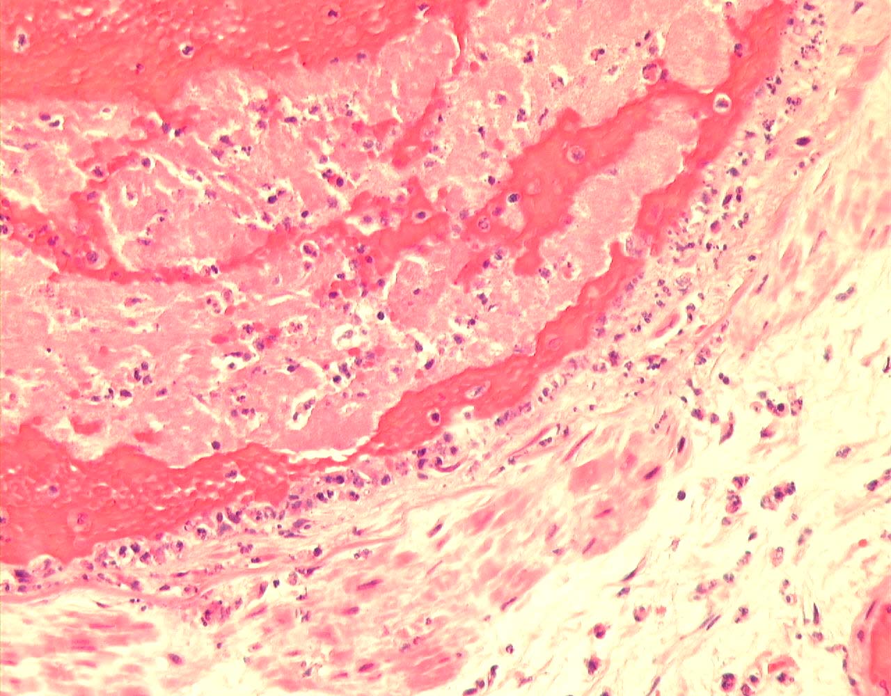
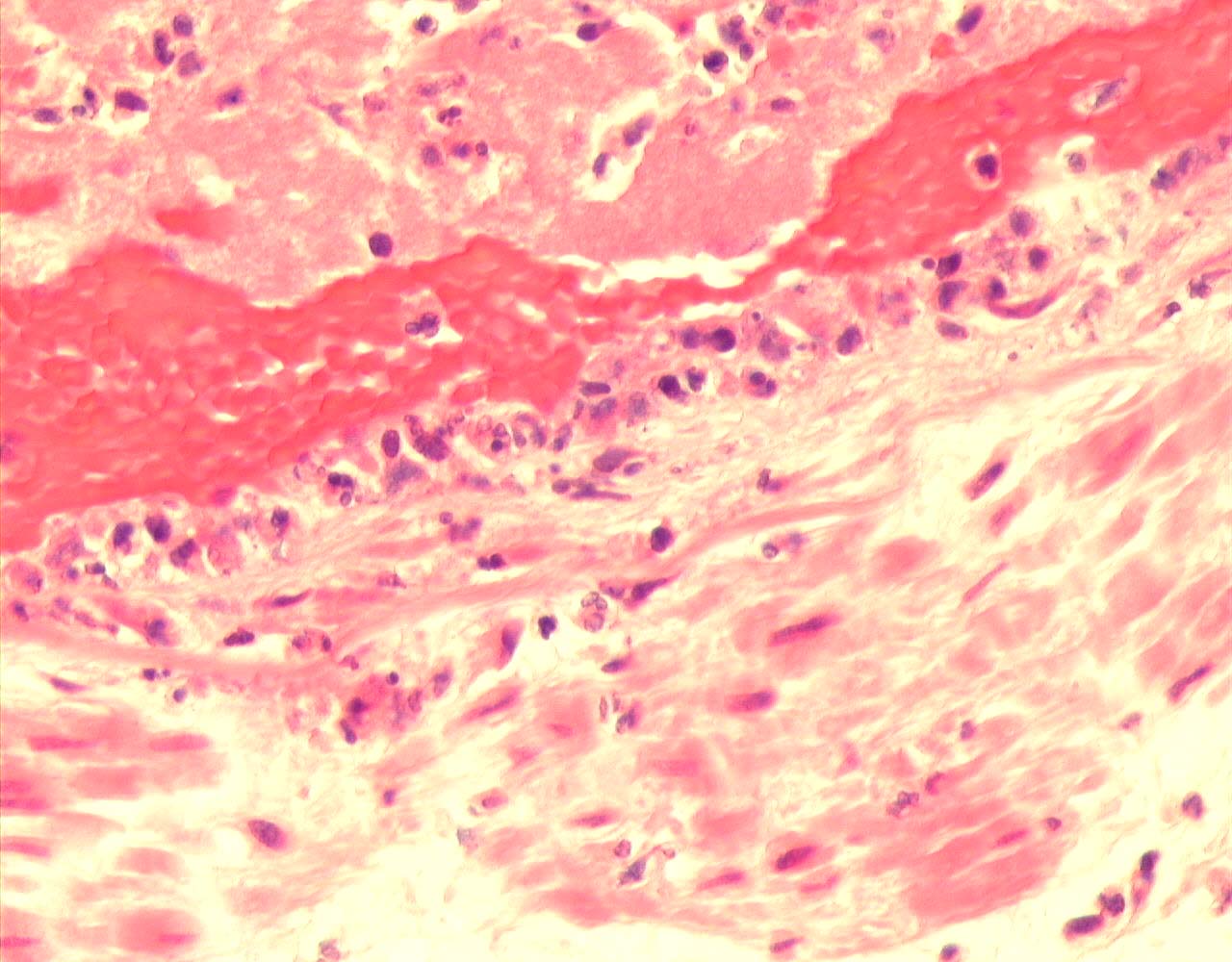
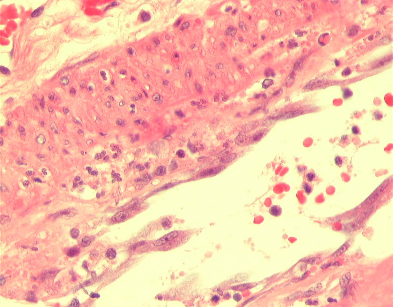
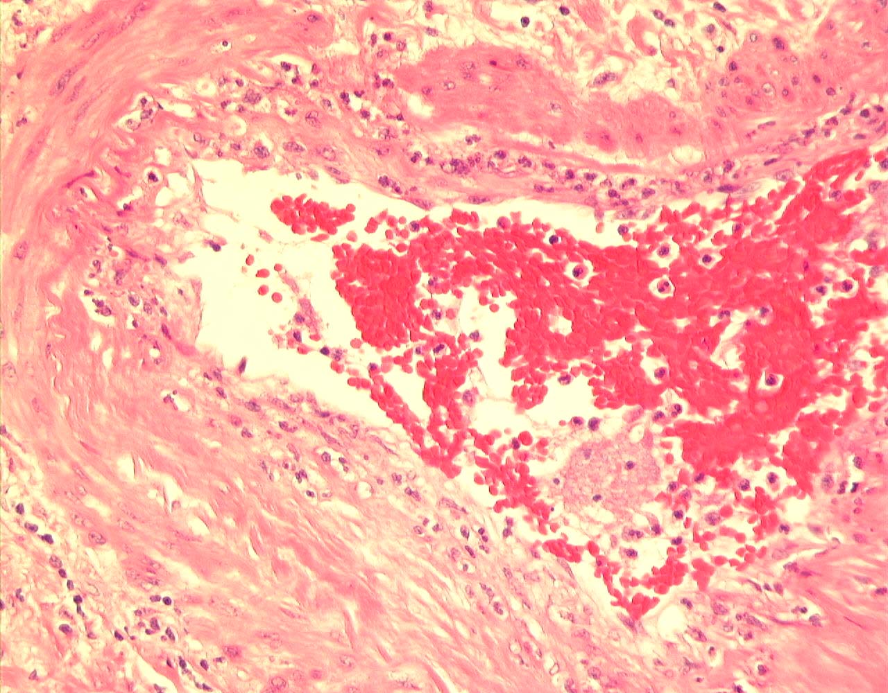
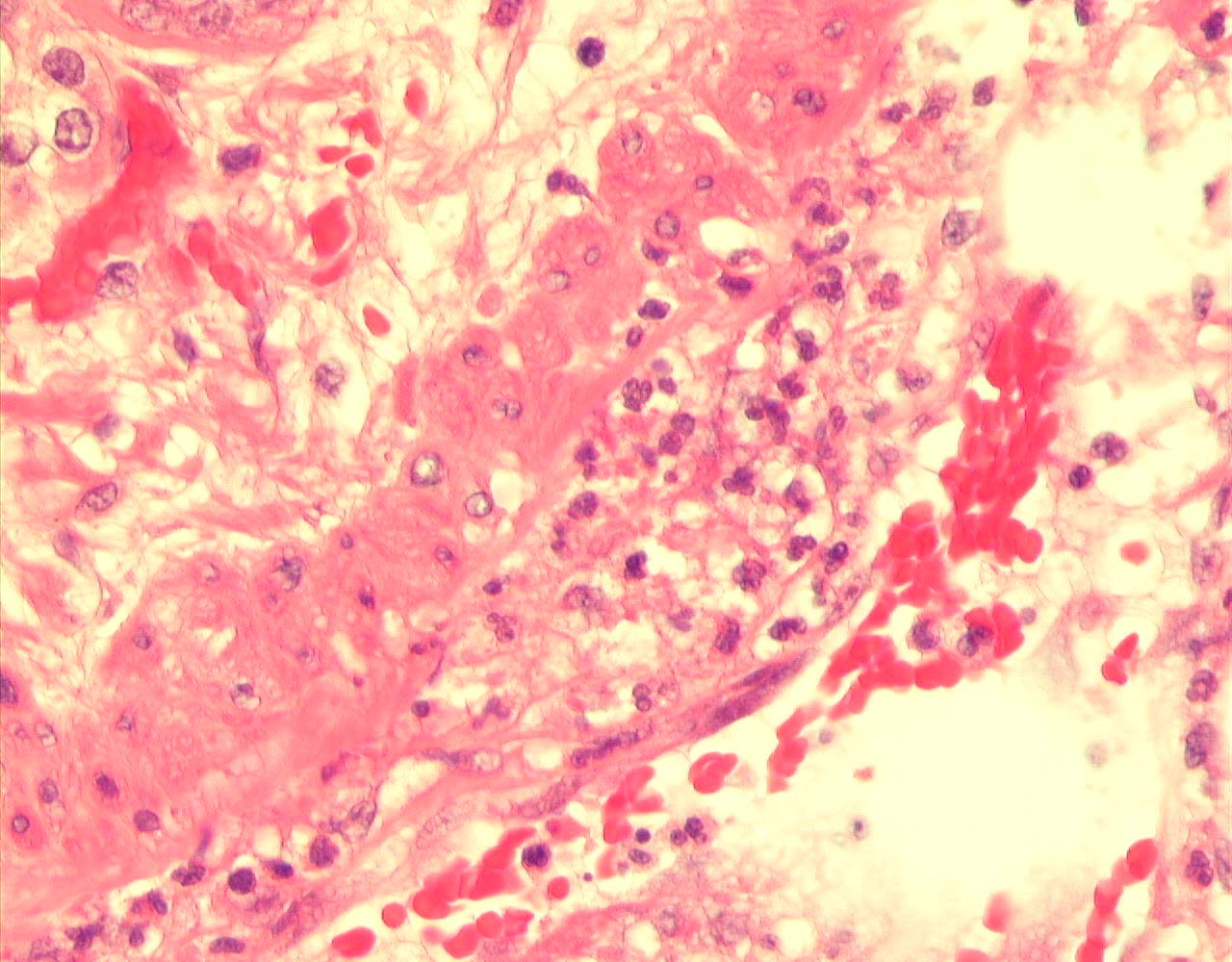
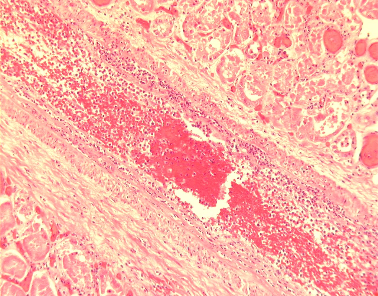
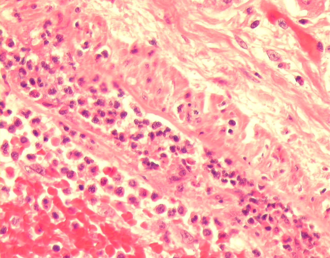
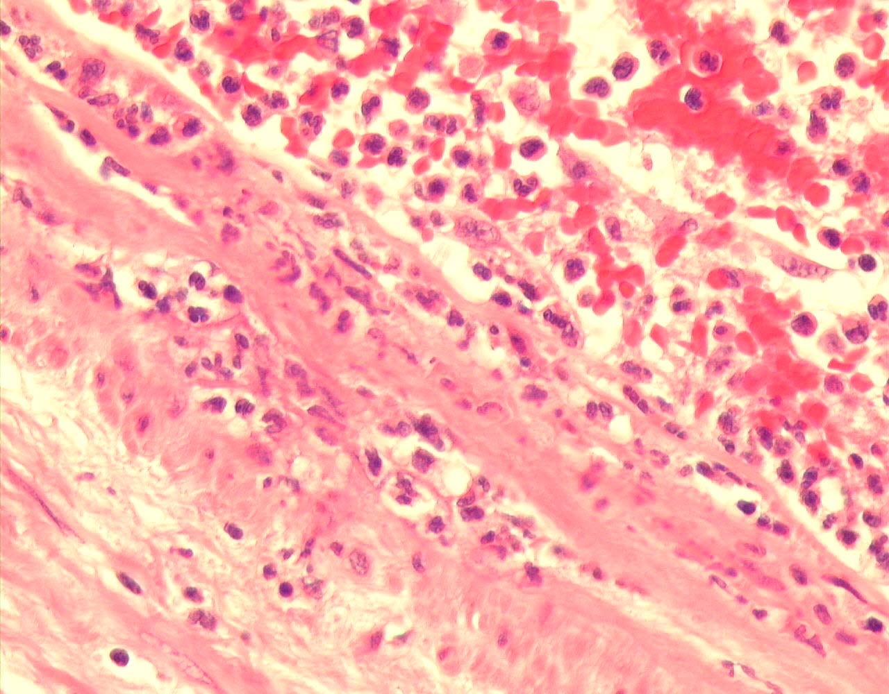
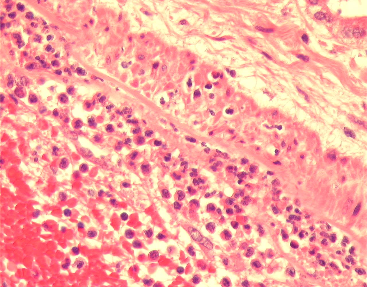
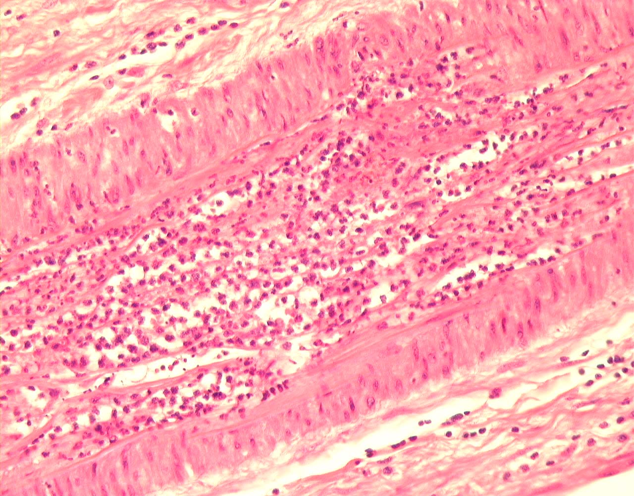
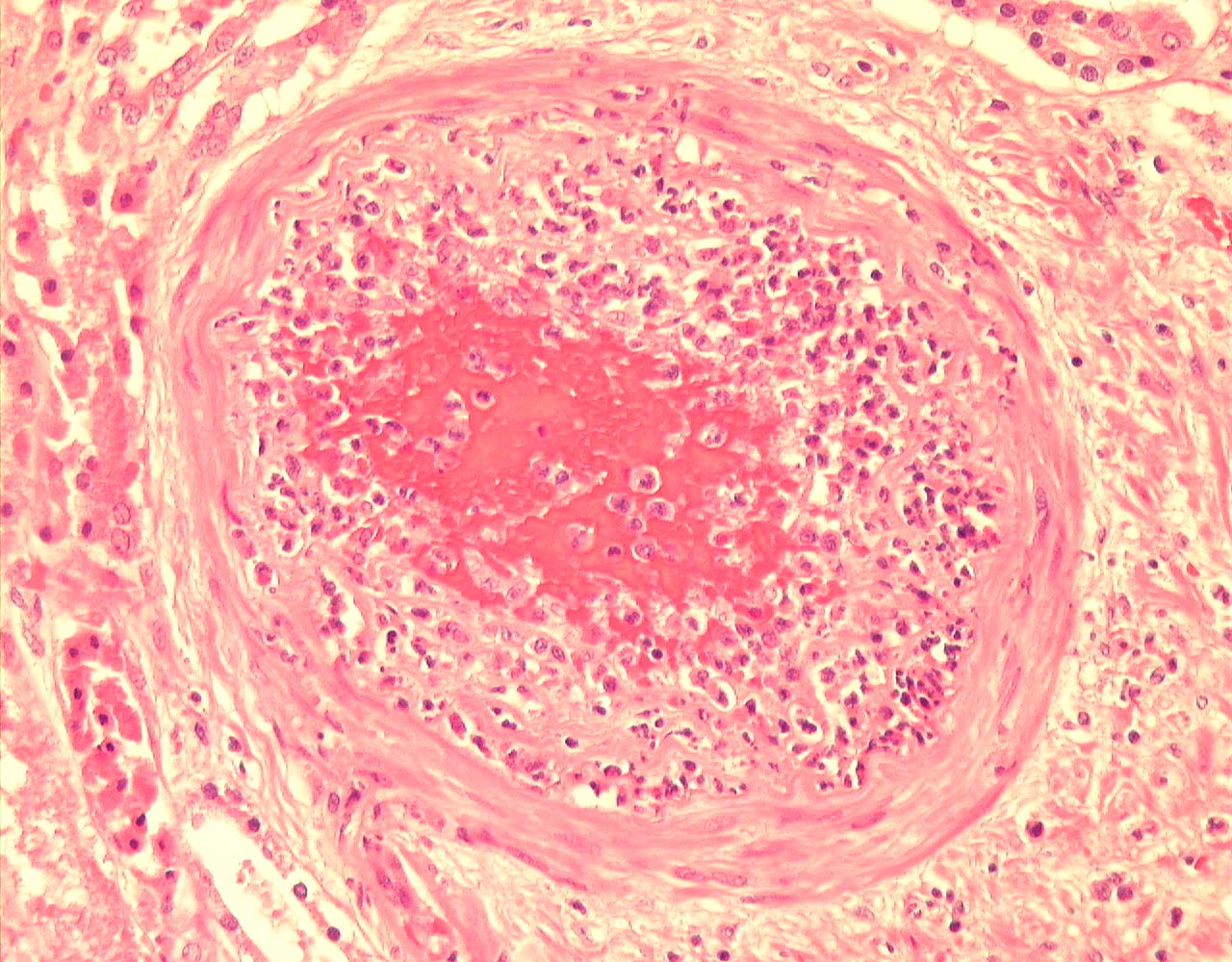
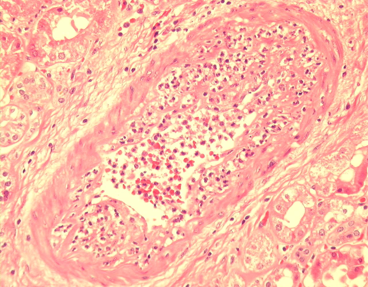
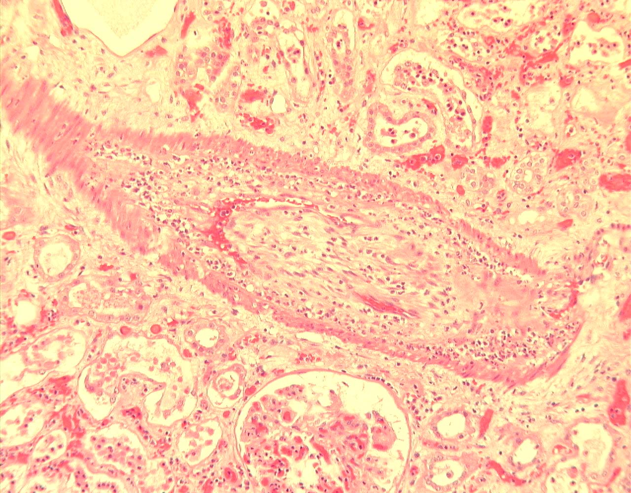
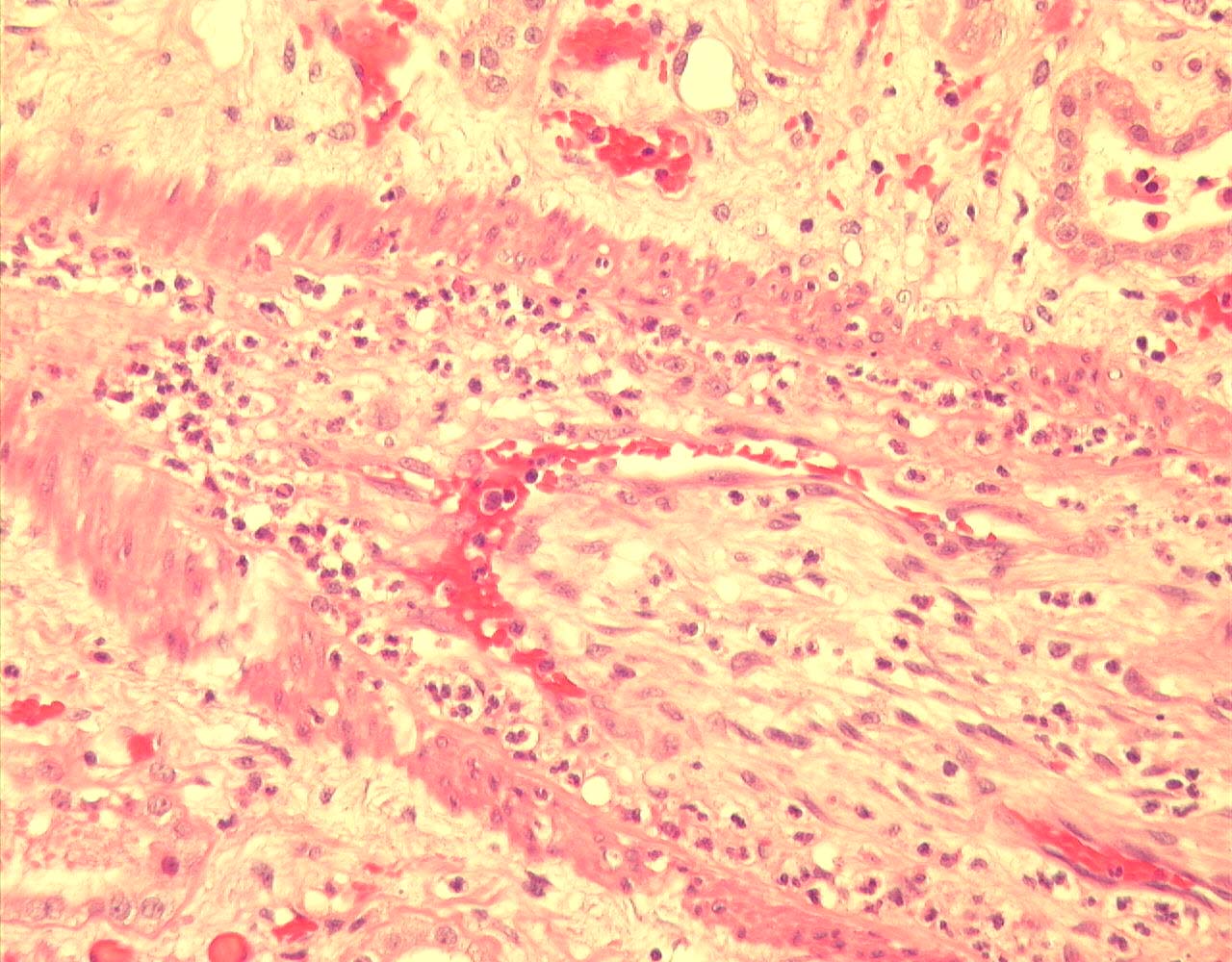
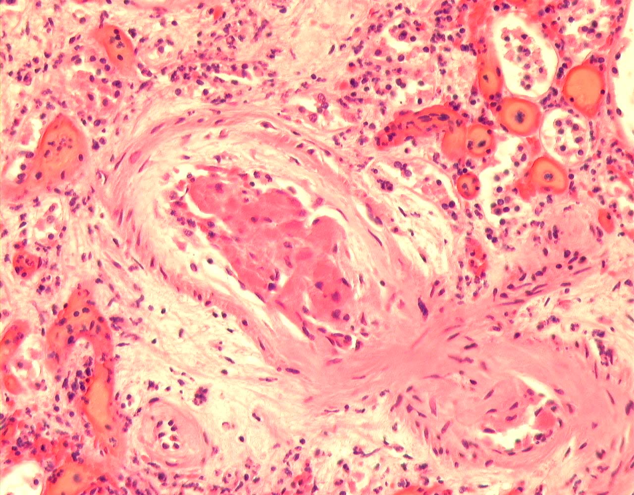
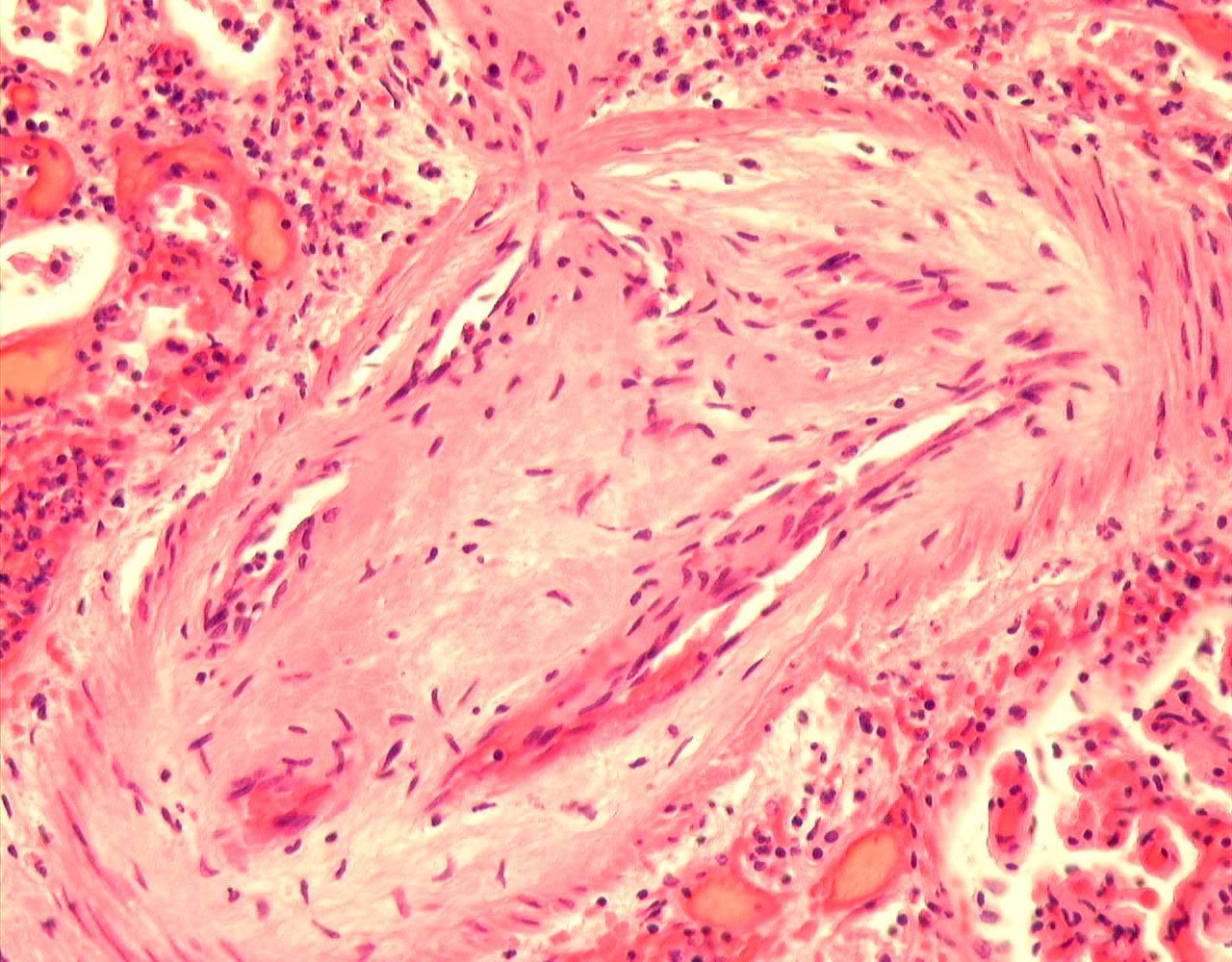
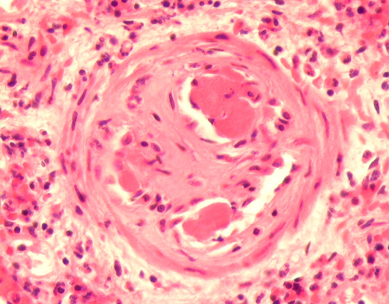
On microscopy, multifocal areas of cortical infarction with an interstitial neutrophil polymorph infiltrate at edge of infarcts.


P02.096_Gibson_1.jpg (205710 bytes) P02.096_Gibson_2.jpg (244247 bytes) P02.096_Gibson_3.jpg (238899 bytes)

Non-infarcted cortex showed congestion, ATN and patchy mild chronic tubulointerstitial damage. There is very mild focal interstitial inflammation away from infarcts, and there is no tubulitis.


P02.096_Gibson_8.jpg (267029 bytes) P02.096_Gibson_17.jpg (248304 bytes)

There are thrombosed arteries at the hilum and in the parenchyma with associated widespread predominantly intimal acute arteritis of small, medium and large vessels.  The arteritis is transmural in some vessels, not all show associated thrombosis, and is characterised by a predominantly neutrophil polymorph infiltrate.  No fibrinoid necrosis of arteries is identified.  Arteries also show mild chronic intimal fibrosis, and some show a more cellular intimal fibrosis and appearances suggesting recanalisation of organised thrombus.

P02.096_Gibson_4.jpg (187053 bytes)


P02.096_Gibson_9.jpg (294188 bytes)


P02.096_Gibson_10.jpg (251543 bytes)


P02.096_Gibson_11.jpg (191377 bytes)


P02.096_Gibson_12.jpg (146705 bytes)


P02.096_Gibson_7.jpg (149772 bytes)


P02.096_Gibson_5.jpg (214677 bytes)


P02.096_Gibson_6.jpg (163919 bytes)


P02.096_Gibson_13.jpg (278410 bytes)


P02.096_Gibson_14.jpg (183572 bytes)


P02.096_Gibson_15.jpg (171377 bytes)


P02.096_Gibson_16.jpg (187166 bytes)


P02.096_Gibson_26.jpg (221502 bytes)


P02.096_Gibson_18.jpg (239148 bytes)


P02.096_Gibson_19.jpg (225610 bytes)


P02.096_Gibson_20.jpg (218565 bytes)


P02.096_Gibson_21.jpg (244896 bytes)


P02.096_Gibson_22.jpg (216853 bytes)


P02.096_Gibson_23.jpg (218010 bytes)


P02.096_Gibson_24.jpg (216199 bytes)


P02.096_Gibson_25.jpg (164422 bytes)


In summary, there is multifocal cortical infarction, arterial thromboses, a polymorph-rich non-necrotising arteritis, ATN and mild chronic allograft nephropathy.

Banff scores: g0,i1,t0,v3,ah0,cg0,mm0,ci1,ct1,cv1

PowerPoint Presentation


(Contact Us)


Copyright © 2000-2007 cyberNephrologyTM All rights reserved. 
Last Modified: Thursday March 08, 2007 05:21:15 PM
info@cybernephrology.org